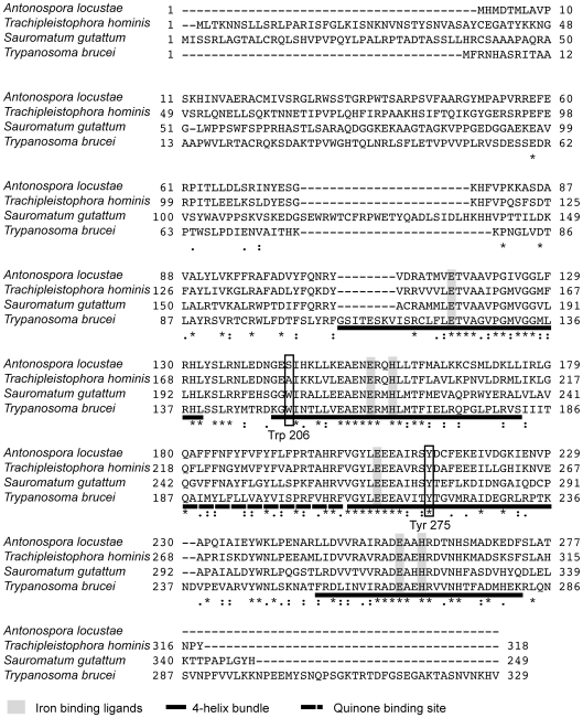
Figure 1. Alignment of A. locustae, T. hominis, S. guttatum and T. brucei AOX sequences.
The four-helix bundles are underlined with a solid line. The putative quinone binding site is underlined with a broken line. Conserved amino acid sites are marked with a star and semi conserved sites are marked with dots.

