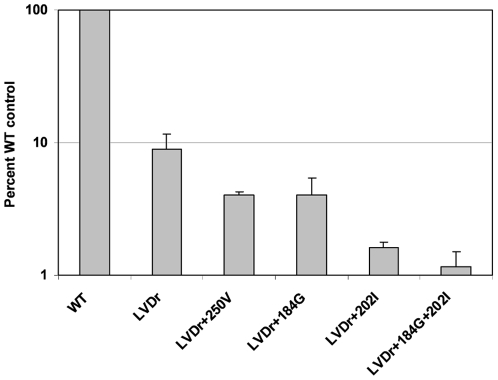
Figure 1. Incorporation of [3H]-ETV into HBV nucleocapsid DNA in culture.
[3H]-ETV was added to cultures of HepG2 cells transfected with an HBV expression construct, as detailed under Materials and Methods. HBV nucleocapsids were isolated from cell lysates, as detailed under Materials and Methods, and radiolabeled HBV DNA was quantified through scintillation counting. The levels of nucleocapsid-associated [3H] from cells grown in [3H]-ETV are presented as percent wildtype control values ± standard deviation. Yields of HBV nucleocapsid DNA were standardized according to real-time PCR quantification of HBV DNA within isolated nucleocapsids, as detailed under Materials and Methods. Similar results were obtained by standardizing total HBV nucleocapsid DNA levels with nucleocapsids from parallel cultures metabolically labeled with [3H]-thymidine (data not shown). WT, wildtype nucleocapsids; LVDr, M204V+L180M substituted HBV nucleocapsids, LVDr+M250V, M204V+L180M+M250V substituted nucleocapsids; LVDr+T184G+S202I, M204V+L180M+T184G+S202I substituted nucleocapsids. HBVs were tested independently 3 to 4 times, except the LVDr+M250V, which was tested twice.

