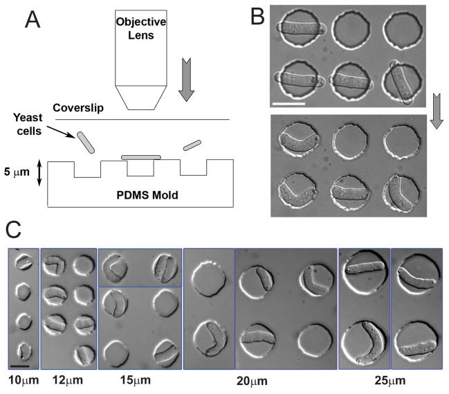
Figure 1. Bending fission yeast cells in situ.
(A) Schematic illustration of the technique used to bend fission yeast cells in situ: cells in liquid media are placed between a PDMS array of microwells and a glass coverslip. The coverslip is subsequently pressurized by overfocusing the objective of an upright microscope. (B) DIC images of the same fission yeast cells before and after pressurizing with the objective. Strain: FC504 (cdc25-22 at 25°C). (C) DIC images of fission yeast cells of varying sizes bent in holes having different diameters (indicated at the bottom). Strains: FC420, FC504. Scale bars: 10μm.

