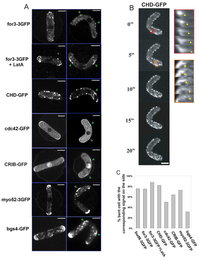
Figure 5. Accumulation of polarity factors and actin assembly at ectopic sites in bent cells.
(A) Localization of the indicated polarity factor fused to GFPs in straight and bent cells, the green arrows point to the sites of recruitment on the sides of the bent cells. Images are maximum confocal projections except for CHD-GFP and cdc42-GFP which are single focal planes. Strains from top to bottom: NM07, NM33, NM59, NM145 (all cdc25-22 at 25°C); FC1371 + HU and NM15 (cdc25-22 at 25°C). (B) Time-lapse imaging of a bent cell expressing an F-actin marker GFP-CHD. Kymographs show that actin filaments are growing from ectopic sites of the cell. The arrowheads indicate the end of the elongating actin cable. Single-plane confocal images are shown. Strain: NM33. (C) Proportion of wildtype bent cells that show a specific location of the indicated factors to the cell’s outer curvature (n≈30 for each condition). Scale bars: 5μm.

