
Figure 5. Cell division is not a requirement for integration and is slowed by integrase protein expression.
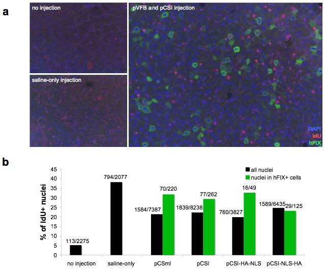
C57BL/6 mice were given 20 μg of pVFB and 20 μg of integrase expression vector (pCSmI, pCSI, pCSI-HA-NLS, or pCSI-NLS-HA) via hydrodynamic injection. Mice were provided with IdU-laced drinking water for one week following hydrodynamic injection. 13 weeks after DNA delivery, livers were fixed and stained for IdU to label cycling cells and hFIX to label cells that were likely to have received integration events. (a) Representative sections are shown for mice given no injection, saline-only injection, or injection of pVFB and pCSI. In many of the hFIX-positive cells, the nuclei do not stain for IdU, indicating that DNA replication did not occur during the week following the procedure. (b) Multiple sections from the injected mice were quantified by counting the number of IdU positive cells within all nuclei (black bars) or the number of IdU positive nuclei within cells that stained positive for hFIX (green bars). The sections chosen deliberately focused on areas that had a high number of hFIX-positive cells, so that a maximum number of hFIX-positive cells could be counted for each group. Consequently, the percentage of cells that were hFIX-positive in Fig. 5b was not representative of the total percentage of hFIX positive cells in the sample. The percentage of IdU positive cells was graphed and the raw data used to calculate that percentage is shown over each bar. All mice used for quantification had the expected serum hFIX level the day following the injection, indicating that the procedure was successful. Hydrodynamic injection without DNA significantly increased proliferation in the liver (Chi-squared test, p<0.0001). There was a decrease in proliferation relative to saline-only injected mice when hFIX and any form of integrase protein were expressed (p<0.0001). The number of IdU positive cells in the population staining positive for hFIX was significantly higher than the overall population for pCSmI (n=5; p<0.0046) and pCSI (n=4; p<0.0369) but not for pCSI-NLS-HA (n=3) or pCSI-HA-NLS (n=2). In cells that were positive for hFIX (green bars), 71% were not replicating their DNA after the hydrodynamic procedure, indicating that φC31 integrase did not require cell division for integration into hepatocyte genomic DNA.