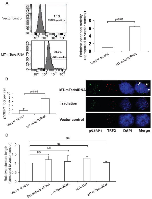
Figure 5. MT-mTer/siRNA induces rapid apoptosis and DNA damage in E4 cells without altering bulk telomere length.
(A) Left: TUNEL assay performed 4 days after lentiviral expression of MT-mTer/siRNA demonstrates brisk apoptotic cell death, 90.7% versus 1.1% with vector control by FACS analysis; shown are representative plots of three different assays with statistical significance (p < 0.01). Right: Caspase assay performed at the same day 4 time point reveals significantly increased caspase activity consistent with apoptosis in cells expressing MT-mTer/siRNA versus control. (B) Left: Quantitation of p53BP1 DNA damage foci in MT-mTer/siRNA or vector control infected cells. Cells were grown on glass cover slips, fixed and stained on day 4 post lentiviral infection, and foci were counted under 63X magnification using a Zeiss Imager.Z1. Right: Representative fluorescence micrographs depicting p53BP1 foci, TRF2, DAPI, and merge. Cells were grown, infected, fixed, and stained as before, then photographed at 63x magnification using a Zeiss LSM 510 confocal scope. MT-mTer/siRNA-infected cells appear to have more p53BP1 DNA damage foci than vector control-infected cells and more co-localization of p53BP1 DNA damage foci with telomeres (TRF2) than irradiated cells (co-localization indicated by arrows, right). (C) Cells infected with lentivirus expressing the various constructs were harvested 3 days after infection and assayed for bulk telomere lengths using RT-PCR (mean of triplicate experiments).

