Abstract
Angioleiomyomas are rare and benign smooth muscle tumors that are infrequently found in the hand. We present a case of angioleiomyoma of the distal ulnar artery that presented with painless mass and gradual enlargement. 3D computed tomography angiography revealed a mass involved with ulnar artery. Surgical excision was performed, and the histology was characteristic of an angioleiomyoma. The patient became asymptomatic after the operation. At 1-year follow-up after the operation, no recurrence has developed. The purpose of this case report is, furthermore, to consider the differential diagnosis in painless masses of the hand.
Keywords: Angioleiomyoma, Ulnar artery, Hand, Tumor
Introduction
Angioleiomyomas are benign, solitary, smooth muscle tumors that can arise anywhere in the body [2, 7, 8]. They originate from the tunica media layer of vessel walls and are uncommon in the hand [3]. The majority of these tumors in the hand had been reported as arising from the veins [5]; only few cases have been reported as arising from the arteries in the hand [1, 2, 11–13, 15]. The rarity of the condition may, in fact, result from incorrect diagnoses. The differential diagnosis of angioleiomyoma of the hand is difficult. Diagnosis is usually made after excision and a histopathologic study of the tumor [3, 8, 13]. We present here a case of angioleiomyoma arising from the distal ulnar artery in the hand and discuss about possible etiology, investigation modalities, differential diagnosis, treatment, and prognosis.
Case Report
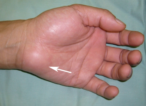
A 37-year-old right-hand-dominant fencer by profession presented with a mass on the right hypothenar region which had been present for several years (Fig. 1). The patient noted that it arose insidiously and reported no history of trauma. He sought treatment as he felt that it had recently increased in size. He denied any pain or recent weight loss, reported no history of cold intolerance, and was working at the time of presentation. On physical exam, there was noted to be a firm mass approximately 3 × 2 cm in size located on the hypothenar eminence without associated muscle atrophy and sensory disturbance. The skin was intact, and there was no erythema. The mass was slightly mobile and minimally tender. The results of preoperative laboratory examinations, including a full blood count with differential cell count and chemistry profile, were within the reference ranges.
Figure 1.
The arrow points to a mass in the center of the hypothenar aspect of right palm.
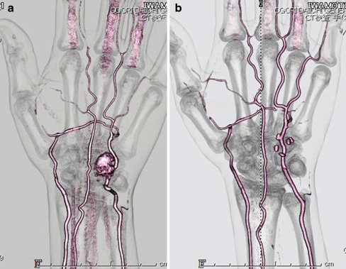
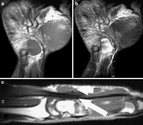
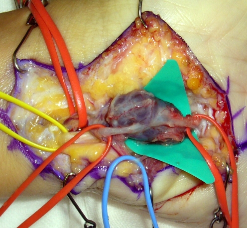
The preliminary differential diagnoses at the time of first presentation to our clinic were ganglion, hemangioma, and lipoma. The conventional radiography disclosed no significant abnormality and the 3D computed tomography angiography (3D CTA) revealed that principal blood supply to this mass came from the distal ulnar artery and a vascular soft tissue mass was presumed (Fig. 2a). A magnetic resonance imaging (MRI) revealed that signal intensity of the longitudinally oriented lesion was predominantly isointense with that of muscle on T1-weighted MRIs and T2-weighted MRIs showed areas of mixed signal intensities that were both hyperintense and isointense to skeletal muscle and also revealed a hypointense rim (Figs. 3a, b). The patient chose to have the mass excised on an elective basis due to its recent increase in size. Excision of the mass was performed under anesthesia with proximal tourniquet control. A 6-cm zigzag skin incision was made on the hypothenar region of the hand. Sharp and blunt dissections were used to dissect the level of the mass. At surgery, a 3 × 2 × 1-cm well-defined mass which was encapsulated and separate from the underlying surrounding tissues or structures was found (Fig. 4). Since the mass was in continuity with the distal ulnar artery, excision required resection segment of the artery followed by an end-to-end microsurgical anastomosis of the ulnar artery. The skin was closed and the hand was placed in a bulky hand dressing.
Figure 2.
a Preoperative 3D CTA of the right hand demonstrates a vascular mass related to ulnar artery. b Postoperative 3D CTA showed intact circulation of ulnar artery after the anastomosis.
Figure 3.
MRI confirmed the presence of the tumor in the subcutaneous tissue. Coronal views: a a T1-weighted image reveals a homogenous well-demarcated soft tissue mass with a signal isotense to muscle. b On a T2-weighted image, there are areas of intermingled hyperintensity and hypointensity inside the mass. Sagittal view: c T1-weighted sagittal image demonstrates the relationship between the soft tissue mass (small arrow) and the underlying flexor tendon sheath (larger arrow).
Figure 4.
The intraoperative clinical picture shows the morphologic features of the tumor.
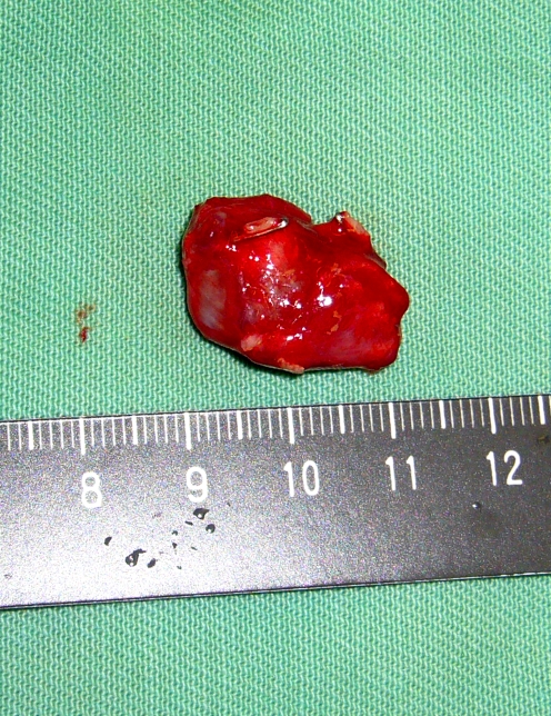
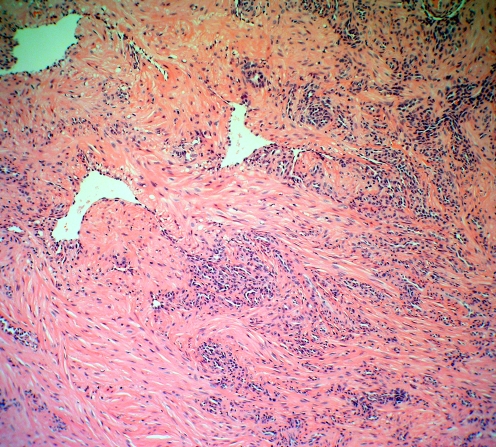
On gross examination, the specimen was firm, rubbery, and hemorrhagic on all sides (Fig. 5). On the histological examination, mass was composed of interlacing and spindle-shaped tumor cell bundles, which were distributed around numerous slit-like vascular channels (Fig. 6). There was regularity of the nuclei but no mitosis of smooth muscle cells and these features confirmed a case of angioleiomyoma. At 2 months follow-up, 3D CTA showed intact circulation of ulnar artery (Fig. 2b) and at the 1-year follow-up evaluation, he had no evidence of recurrence of the mass, no pain and any cold intolerance.
Figure 5.
The macroscopic appearance of the tumor is shown.
Figure 6.
Microscopic findings show numerous vascular channels embedded within a smooth muscle component (stain, H&E; magnification, ×100).
Discussion
Angioleiomyoma is a relatively nonaggressive tumor of smooth muscle originating from the blood vessels, comprising approximately 4.4% of all benign soft tissue tumors [7]. The exact etiology of angioleiomyomas is unknown. Minor trauma, hamartoma changes, venous stasis, and hormonal imbalance have been proposed as etiological features [4, 7, 14]. An arteriovenous malformation has also been cited in the literature [6]. In our case, patient is a fencer by profession, and we assume that repeated use of foil may have caused the injury to ulnar artery in the hypothenar region.
Clinically, the most common complaint associated with a vascular leiomyoma of the hand is painless swelling or slightly tender tumor mass [2, 10]. Growth is usually slow and it may be years before a patient seeks medical evaluation. Most angioleiomyoma located in the hand do not show direct relationship to arteries, and only few tumors involving arteries have been reported so far [1, 2, 11–13, 15]. There are no specific imaging techniques for angioleiomyoma, but there are reports of 3D CTA imaging [15] and 3D MRI [12] being performed before surgery to help clinicians understand exactly the relationship between a angioleiomyoma and the involved arteries. In the current case, preoperative 3D CTA clearly showed the mass involving the ulnar artery and MRI defined its relationship with adjacent structures (Fig. 3c), allowing better surgical planning.
Angioleiomyoma does not have distinctive clinical presentation or radiographic features and is rarely diagnosed before surgery [2, 10]. The differential diagnosis can include true or false aneurysm, hemangioma, lipoma, ganglion, pigmented villonodular synovitis, giant cell tumor of a tendon sheath, and neurofibroma [2, 3, 8]. Since angioleiomyomas are similar in clinical presentation and painless, familiar to other types of tumors of the hand, the clinical diagnosis is often difficult to make and, thus, histopathologic examination is required for a definitive diagnosis of angioleiomyoma.
The usual treatment for patients having symptoms similar to those in our case is simple excision of the mass and ligation of the feeder vessels [12]. Adequate collateral circulation should be verified both before and during the operation. If collateral flow is not adequate, vascular repair or grafting may be necessary. In our case, even though collaterals were adequate, we did end-to-end anastomosis of the ulnar artery because ulnar artery is the dominant artery of the hand and maintaining adequate circulation in the hand will prevent any vascular problems as well as cold intolerance. During surgery, careful dissection to prevent damage to surrounding tendons, vessels, and nerves is recommended. Most hand angioleiomyomas are benign but malignant tumors have been reported [9]. If resection is complete, the chance of recurrence is minimal and the prognosis is excellent.
In conclusion, angioleiomyoma is a rare tumor of the hand. Its diagnosis must be considered when a patient presents with an isolated painless mass with a slow growth. The prognosis is good after surgical excision without risk of recurrence.
Footnotes
All the authors in this article equally contributed, and neither the authors nor the related institution has received any financial benefit.
References
- 1.Botte MJ, Silver MA. Leiomyoma of a digital artery. Clin Orthop Relat Res. 1990;260:259–262. [PubMed] [Google Scholar]
- 2.Callé SC, Eaton RG, Littler JW. Vascular leiomyomas in the hand. J Hand Surg [Am] 1994;19:281–286. doi: 10.1016/0363-5023(94)90020-5. [DOI] [PubMed] [Google Scholar]
- 3.Duhig J, Ayer J. Vascular leiomyoma: a study of 61 cases. Arch Pathol. 1959;68:424–430. [PubMed] [Google Scholar]
- 4.Duinslaeger L, Viererdeels T, Wylock P. Vascular leiomyoma in the hand. J Hand Surg [Am] 1987;12:624–627. doi: 10.1016/s0363-5023(87)80223-x. [DOI] [PubMed] [Google Scholar]
- 5.Enzinger FM, Weiss SW. Benign tumors of smooth muscle. In: Weiss SW, Goldblum JR, editors. Soft tissue tumors. 4. St. Louis: Mosby; 1995. pp. 695–726. [Google Scholar]
- 6.Fox S, Heryet A, Khong T. Angioleiomyomas: an immunohistological study. Histopathology. 1990;16:495–496. doi: 10.1111/j.1365-2559.1990.tb01074.x. [DOI] [PubMed] [Google Scholar]
- 7.Hachisuga K, Hasimoto H, Enjoji M. Angioleiomyoma, a clinicopathologic reappraisal of 562 cases. Cancer. 1984;54:126–130. doi: 10.1002/1097-0142(19840701)54:1<126::AID-CNCR2820540125>3.0.CO;2-F. [DOI] [PubMed] [Google Scholar]
- 8.Herren DB, Zimmermann A, Buchler U. Vascular leiomyoma in an index finger undergoing malignant transformation. J Hand Surg (Br) 1995;20:484–487. doi: 10.1016/s0266-7681(05)80158-5. [DOI] [PubMed] [Google Scholar]
- 9.Hirata H, Kusuzaki K, Fukutome K, et al. A hand mass that became painful 13 years after onset. Clin Orthop Relat Res. 2005;433:265–270. doi: 10.1097/01.blo.0000150575.90588.21. [DOI] [PubMed] [Google Scholar]
- 10.Lawson GM, Salter DM, Hooper G. Angioleiomyomas of the hand. A report of 14 cases. J Hand Surg (Br) 1995;20:479–483. doi: 10.1016/s0266-7681(05)80157-3. [DOI] [PubMed] [Google Scholar]
- 11.Miyamoto W, Yamamoto S, Kii R, et al. Vascular leiomyoma resulting in ulnar neuropathy: case report. J Hand Surg [Am] 2008;33:1868–1870. doi: 10.1016/j.jhsa.2008.07.008. [DOI] [PubMed] [Google Scholar]
- 12.Nagata S, Nishimura H, Uchida M, et al. Giant angioleiomyoma in extremity: report of two cases. Magn Reson Med Sci. 2006;5:113–118. doi: 10.2463/mrms.5.113. [DOI] [PubMed] [Google Scholar]
- 13.Ramesh P, Annapureddy SR, Khan F, et al. Angioleiomyoma: a clinical, pathological and radiological review. Int J Clin Pract. 2004;58:587–591. doi: 10.1111/j.1368-5031.2004.00085.x. [DOI] [PubMed] [Google Scholar]
- 14.Robinson SC, Kalish RJ. Leiomyoma in the hand: a case report. Clin Orthop. 1990;255:121–123. [PubMed] [Google Scholar]
- 15.Yagi K, Hamada Y, Yasui N. A leiomyoma arising from the deep palmar arterial arch. J Hand Surg [Br] 2006;31:680–682. doi: 10.1016/j.jhsb.2006.08.006. [DOI] [PubMed] [Google Scholar]


