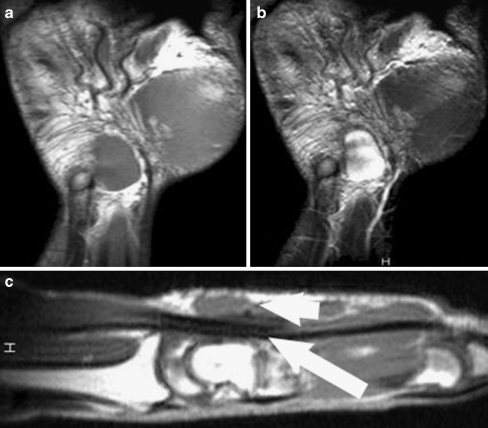
Figure 3.
MRI confirmed the presence of the tumor in the subcutaneous tissue. Coronal views: a a T1-weighted image reveals a homogenous well-demarcated soft tissue mass with a signal isotense to muscle. b On a T2-weighted image, there are areas of intermingled hyperintensity and hypointensity inside the mass. Sagittal view: c T1-weighted sagittal image demonstrates the relationship between the soft tissue mass (small arrow) and the underlying flexor tendon sheath (larger arrow).

