
Figure 1.
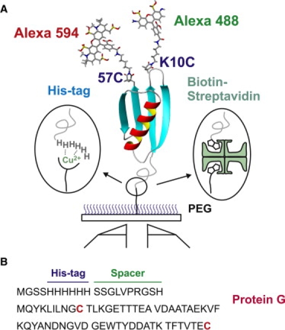
Design of the protein and immobilization of molecules. (A) Immobilization of dye-labeled proteins via his-tag–Cu2+ or biotin (protein)–streptavidin–biotin (surface) linkages on a polyethylene-glycol–coated glass surface. Donor (Alexa 488) and acceptor (Alexa 594) dyes are labeled at the cysteine residues at positions 10 and 57 of protein GB1 (His-GB1K10C/C57). The two isomers, donor10/acceptor57 and donor57/acceptor10, could not be separated in the purification. (B) Amino acid sequences of protein, spacer, and his-tag. Dyes are labeled on cysteine residues (red). The N-terminus of protein GB1 is tethered to six histidine residues with a 10-residue spacer.