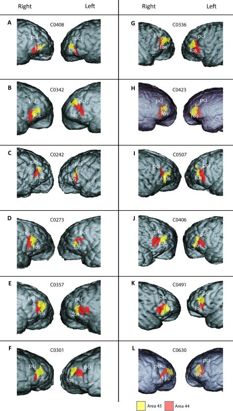
Figure 3.
Reconstructed lateral views of the magnetic resonance images of both hemispheres in 12 chimpanzees. The extent of areas 44 and 45 on the lateral surface of the brain are shown in red and yellow, respectively. The position of areas 44 and 45 where they lie within sulci is not visible (A: C0408, B: C0342, C: C0242, D: C0273, E: C0357, F: C0301, G: C0336, H: C0423, I: C0507, J: C0406, K: C0491, and L: C0630). Abbreviations: fos = fronto-orbital sulcus; pci = inferior precentral sulcus.

