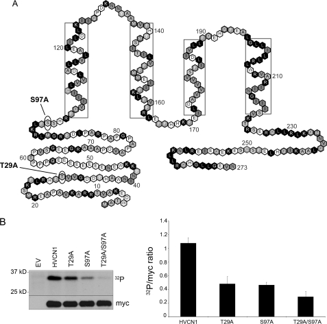
FIGURE 1.
Identification of candidate phosphorylation sites in the human proton channel. A, location of the two putative PKC-δ phosphorylation sites in the sequence of HV1. Both predicted PKC-δ sites are in the N terminus, with Ser97 near the membrane boundary. Boxed residues indicate helical transmembrane regions (37). B, phosphorylation of HV1 WT, T29A, S97A, and T29A/S97A mutants in the presence of recombinant PKC-δ and [γ-32P]ATP in an in vitro kinase assay. PKC-δ was activated with 1 μm PMA. The myc immunoblot indicates loading. EV indicates empty vector control. The graph on the right-hand side represents densitometric analysis of the 32P band versus loading control of 3–4 separate experiments (p < 0.05 for each mutant versus WT by Student's t test). Error bars indicate S.E.

