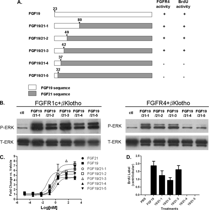
FIGURE 4.
FGF19/21 chimeric proteins. A, schematic diagram showing chimeric proteins between FGF19 and FGF21. The numbers for the last residue from N-terminal FGF19 sequences in each chimeric construct are shown. B, L6 cells were transfected with FGFR1c or FGFR4 and with β-Klotho. Following overnight serum starvation, cells were stimulated with vehicle or with 50 nm of recombinant FGF19, FGF21, or chimeric proteins for 10 min and snap frozen in liquid nitrogen. Cell lysates were prepared for Western blot using antibodies against phosphorylated ERK1/2 (p-ERK) or total ERK1/2 (T-ERK). C, differentiated 3T3-L1 adipocytes were incubated for 72 h with recombinant FGF19, FGF21 or chimeric proteins and assayed for glucose uptake. D, semiquantitative analysis of BrdU immunostaining of livers from female FVB mice treated for 6 days with PBS, 2 mg/kg/day recombinant FGF19 or 2 mg/kg/day chimeric proteins. The scores assigned to BrdU incorporation for these animals were based on a semiquantitative scale described under “Experimental Procedures”. The BrdU immunostaining of livers were showed in supplemental Fig. S3. Solid bars represent group mean score with S.E. (n = 8 for each group).

