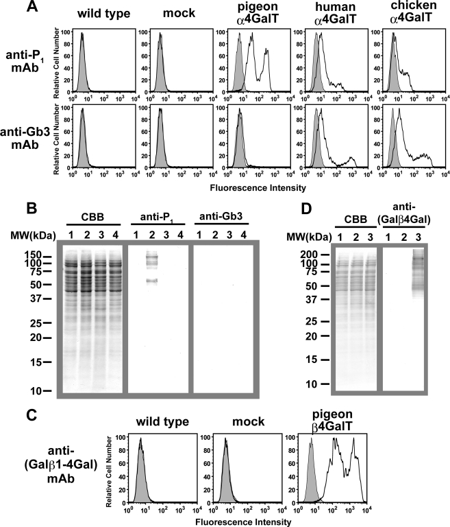
FIGURE 5.
Expression of α4GalT(Gal) or β4GalT(Gal) in 293T cells. A and B, detection of P1 antigen and Gb3 on cells transfected with pigeon, human, or chicken α4GalT(Gal) DNA. Wild-type and 293T cells transfected with mock, pigeon, human, or chicken α4GalT(Gal) DNA were analyzed with FACS (A) or Western blotting (B). A, cell surfaces were stained with anti-P1 mAb or anti-Gb3 mAb, and fluorescein isothiocyanate-goat anti-mouse IgM was used as the secondary antibody. Histograms in gray indicate the results in the absence of primary antibodies. B, Western blotting analysis of cell extracts from cells transfected with mock (lane 1), pigeon (lane 2), human (lane 3), or chicken (lane 4) α4GalT(Gal) DNA. Proteins blotted onto polyvinylidene difluoride membranes were stained with Coomassie Brilliant Blue R-250 (CBB), anti-P1 mAb, or anti-Gb3 mAb, as indicated. C and D, detection of Galβ1–4Gal on cells transfected with pigeon β4GalT(Gal) cDNA. Wild-type and 293T cells transfected with mock or pigeon β4GalT(Gal) cDNA were analyzed with FACS (C) or Western blotting (D). C, cell surfaces were stained with anti-(Galβ1–4Gal) mAb, and fluorescein isothiocyanate-goat anti-mouse IgG was used as the secondary antibody. Histograms in gray indicate the results in the absence of primary antibodies. D, Western blotting analysis of cell extracts from cells transfected with wild-type (lane 1), mock (lane 2), or pigeon β4GalT(Gal) (lane 3) cDNA. Proteins blotted onto polyvinylidene difluoride membranes were stained with Coomassie Brilliant Blue R-250 or anti-(Galβ1–4Gal) mAb.

