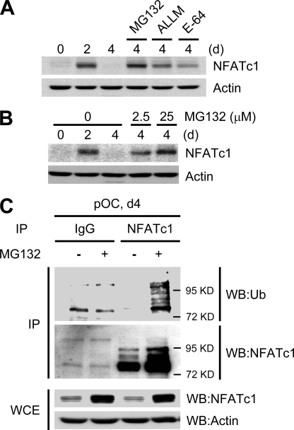
FIGURE 2.
Ubiquitin-mediated NFATc1 degradation. A–C, BMMs were cultured with M-CSF and RANKL for the indicated times. A, the proteasome inhibitor MG132 (25 μm), the calpain inhibitor ALLM (50 μm), or the cysteine inhibitor E-64 (50 μm) were added 6 h before cell lysis on day 4 as indicated. B, various concentrations of MG132 were added 6 h prior to cell lysis on day 4 as indicated. In A and B, lysates were immunoblotted with NFATc1 and actin antibodies. C, BMMs were cultured with M-CSF and RANKL for 4 days. MG132 (25 μm) was added 4 h before cell lysis on day 4 as indicated. Whole cell lysates (WCE) from mature osteoclasts were immunoprecipitated with anti-NFATc1 or IgG control antibodies; immunoprecipitates (IP) were probed with anti-ubiquitin (Ub, upper panel) or anti-NFATc1 antibodies (middle panel). Whole cell extracts were probed with anti-NFATc1 or anti-actin (control) antibodies (lower panel). WB, Western blot.

