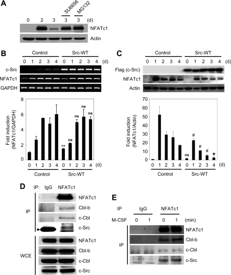
FIGURE 4.
Src-Cbl proteins are involved in down-regulation of NFATc1 protein. A, BMMs were cultured with M-CSF and RANKL for the indicated times. The Src kinase inhibitor SU6656 (2 μm) or MG132 (25 μm) was added 8 h before cell lysis. Lysates were probed with NFATc1 and actin antibodies. B and C, BMMs were transduced with pMX-IRES-EGFP (Control) or c-Src-WT retrovirus (Src-WT) and cultured with M-CSF and RANKL for the indicated times. B, reverse transcription-PCR (upper panel) and real-time PCR (lower panel) were performed to detect c-Src, NFATc1, and glyceraldehyde-3-phosphate dehydrogenase (GAPDH). C, lysates were immunoblotted with FLAG (for overexpressed c-Src), NFATc1, and actin antibodies (upper panel). The relative amounts of NFATc1 are shown in the lower panel. B and C, ns, not significant, #, p < 0.05, *, p < 0.01, **, p < 0.001 versus positive control. Data are expressed as mean ± S.D. of triplicate samples. Results presented are representative of three independent sets of similar experiments. D, lysates from preosteoclasts were immunoprecipitated (IP) with anti-NFATc1 or control IgG antibodies. Immunoprecipitated samples (upper panel) or whole cell lysates (WCE; lower panel) were subjected to Western blotting for detection of NFATc1, Cbl-b, c-Cbl, and c-Src. The arrow indicates the IgG band. E, preosteoclasts were stimulated with M-CSF for the indicated times. Lysates from preosteoclasts were immunoprecipitated with anti-NFATc1 or control IgG antibodies. Immunoprecipitated samples were subjected to Western blot analysis for detection of NFATc1, Cbl-b, c-Cbl, and c-Src.

