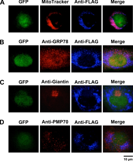
FIGURE 2.
Intracellular localization of human AGXT2 in HUVEC. HUVEC were infected with AdAGXT2FLAG and stained with anti-FLAG antibodies and either: A, MitoTracker; B, anti-GRP78 (a marker of endoplasmic reticulum); C, anti-giantin (a marker of the Golgi); or D, anti-PMP70 (a marker of peroxisomes). Fluorescence confocal microscopy was performed to detect GFP (green), MitoTracker (red), GRP78 (red), giantin (red), PMP70 (red), FLAG (blue), or a merged image of multiple markers (Merge).

