FIGURE 4.
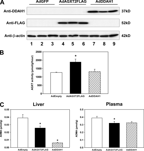
Expression of human AGXT2 in mice. A, C57BL/6 mice were injected retro-orbitally with AdGFP (lanes 1–3), AdAGXT2FLAG (lanes 4–6), or AdDDAH1 (lanes 7–9). Four days after injection, the livers were harvested and subjected to immunoblotting with antibodies to DDAH1, FLAG, or β-actin. Three separate mice were injected with each adenovirus. B, total AGXT activity of liver lysates prepared 4 days after injection of AdEmpty (n = 4), AdAGXT2FLAG (n = 4), or AdDDAH1 (n = 4). C, levels of ADMA in the liver and plasma 4 days after injection of AdEmpty (n = 10 for liver and n = 25 for plasma), AdAGXT2FLAG (n = 10 for liver and n = 25 for plasma), or AdDDAH1 (n = 5 for liver and n = 5 for plasma). Values are mean ± S.E. *, p < 0.05 compared with mice infected with AdEmpty.

