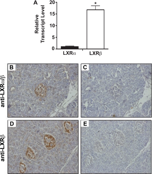
FIGURE 1.
Distribution of LXRs in human islets. A, qRT-PCR was performed using total mRNA isolated from primary human islets maintained in 11 mm glucose. LXR mRNA levels were normalized to β-actin mRNA levels. All data are shown as the mean ± S.E. (n = 3). *, data indicate statistical difference (p < 0.05) compared with LXRα mRNA level. Panels B–E, human pancreas sections were immunostained with anti-LXRα/β antibody (B) or anti-LXRα/β antibody plus blocking peptide for LXRα/β (C). Human pancreas sections were immunostained with anti-LXRβ antibody (D) or anti-LXRβ antibody plus blocking peptide for LXRβ (E). Antibody binding was revealed using peroxidase-conjugated secondary IgG and diaminobenzidine tetrahydrochloride substrate to yield a brown signal.

