FIGURE 7.
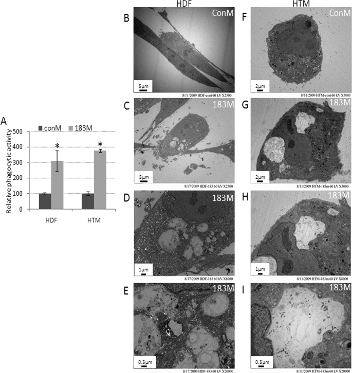
Panel A, increase in phagocytic activity and cell vacuolization induced by miR-183 in HDF and HTM cells. Normal HDF and HTM cells were transfected with 183M or ConM. Two days post-transfection, HDF cells were incubated with collagen-coated fluorescence beads and HTM cells with pHRodo E. coli overnight. Cells were then collected and analyzed by flow cytometry in the FL1 (fluorescence isothiocyante) channel (collagen-coated fluorescence beads) or FL2 (561 nm-laser) channel (pHRodo E. coli). Data represent the change in phagocytic activity in cells transfected with 183M compared with control cultures transfected with ConM ± S.D. (n = 3–5, *, p < 0.05, Mann-Whitney U test). Panels B–I show representative electron microscopy images of increased vacuolization in HDF and HTM cells 3 days after transfection with 183M compared with cells transfected with ConM. The images (×2,500) were recorded in both ConM- (B) and 183M (C)-transfected cells. The images (×5,000) were recorded in both ConM- (F) and 183M (G)-transfected cells. Higher magnification images (×8,000 and 20,000) of vesicles present in cells transfected with 183M are also shown (D and E, HDF; H and I, HTM). The figures are representative results from three independent experiments. Error bars are S.D.

