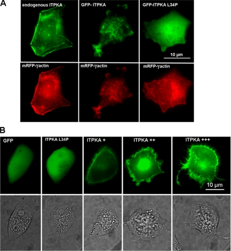
FIGURE 2.
Effects of ITPKA on formation of F-actin-based cell protrusions. A, to analyze co-localization between endogenous ITPKA and F-actin, H1299 cells were transfected with mRFP-γ actin (red, left panel) and stained with an anti-ITPKA antibody (green, left panel). In addition, H1299 cells were co-transfected with GFP-ITPKA (green, middle panel) and mRFP-γ-actin (red, middle panel) fusion proteins. Right panel, actin binding of ITPKA was blocked by L34P (25), and cells were co-transfected with GFP-ITPKA L34P (green) and mRFP-γ-actin (red). Fluorescence of the fusion proteins was monitored by confocal fluorescence microscopy. B, cells were transfected with GFP, GFP-ITPKA L34P, and with GST-ITPKA. After 16 h of incubation, the cells were washed and covered with PBS. Viable cells were monitored by light and by fluorescence microscopy. The protrusions were determined from cells expressing high levels of GFP and GFP-ITPKA L34P and low (+), intermediate (++), or high (+++) levels of GFP-ITPKA (see Table 1).

