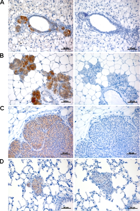
FIGURE 8.
ITPKA expression in highly invasive mouse tumors. Paraffin sections of Balb-neuT tissue were stained for ITPKA (left panels), whereas normal goat IgG was used as a negative control (right panels). Shown are sections of breast tissues of 5-week-old (A), 11-week-old (B), 20-week-old mice (C), and lung metastasis from 24-week-old mice (D). Scale bars, 50 μm.

