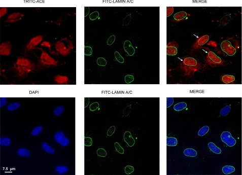
FIGURE 7.
Localization in the nucleus and nuclear envelope. SMC were grown in slide chambers, incubated with TRITC-ACE, and processed for immunofluorescence detection as described under “Experimental Procedures.” Arrows indicate the areas in which lamin A/C (green) co-localize with TRITC-ACE (red). DAPI-stained nuclear DNA (blue) and lamin A/C do not colocalize.

