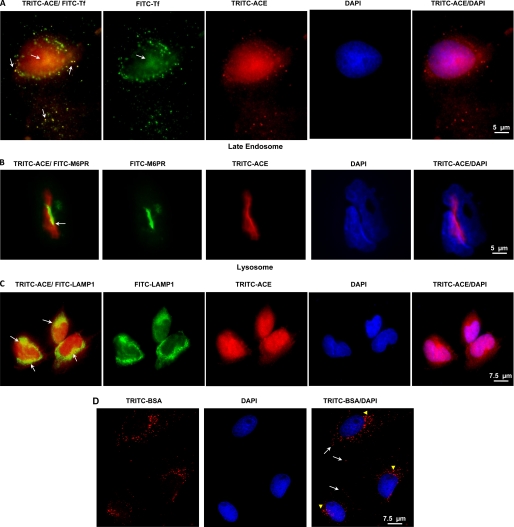
FIGURE 9.
ACE in endosomal compartments and lysosome of SMC. A, SMC monolayers incubated with 10 nm TRITC-ACE (red) and 10 nm FITC-transferrin (FITC-Tf) (green) for 120 min. Cells were also incubated for 120 with 10 nm TRITC-ACE and then with anti-mannose 6-phosphate receptor (M6PR) (B) or anti-LAMP1 (C). Arrows point to ACE localization in the indicated compartment. D, cells were incubated with 10 nm TRITC-BSA as described above for TRITC-ACE. Internalized TRITC-BSA occupied the early endosome (arrows) and late endosome/lysosome (arrowheads) but not the nuclei. Indirect fluorescent microscopy analysis was done as described under “Experimental Procedures.” Scale bar is at the lower right hand corner.

