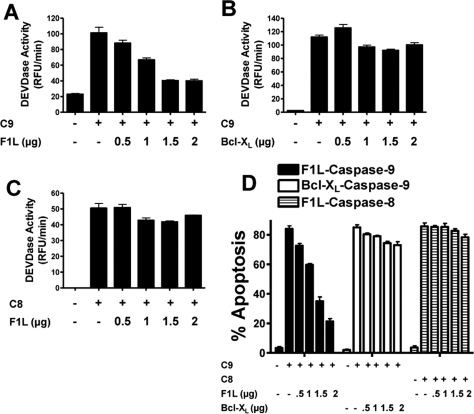
FIGURE 6.
F1L suppresses caspase-9-induced apoptosis. A–C, HEK293T cells were transfected with various amounts of plasmids (pcDNA3 alone (−)) encoding full-length FLAG-F1L or Bcl-XL (0–2.0 μg) with or without FLAG-caspase-9 (0.5 μg (A and B)) or caspase-8 (1 μg (C)) plasmid while maintaining total DNA constant at 3 μg by the addition of pcDNA3. Cell lysates were prepared 20 h after transfection, normalized for protein content (10 μg), and incubated with the caspase-3/-7 substrate Ac-DEVD-AFC. Enzyme activity was determined by the generation of fluorescent AFC product, and Vmax was calculated (mean ± S.D.; n = 3). D, HEK293T cells were transfected with various amounts of plasmids encoding GFP (−), GFP-F1L, or Bcl-XL (0–2.0 μg) with or without FLAG-caspase-9 (0.5 μg) or caspase-8 (1 μg) plasmid while maintaining total DNA constant at 3 μg by the addition of pcDNA3. At 20 h post-transfection, both floating and adherent cells were collected, fixed, and stained with 0.1 μg/ml DAPI. The percentages of apoptotic cells were determined by counting the GFP-positive cells having nuclear fragmentation and/or chromatin condensation (mean ± S.D.; n = 3). RFU, relative fluorescence units.

