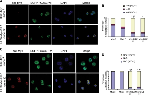
FIGURE 7.
GILZ expression provokes FOXO3-WT and FOXO3-TM accumulation within the cytoplasm. HL-60-Myc and HL-60-Myc-GILZ clones were transiently transfected with 10 μg of pEGFP-FOXO3-WT plasmid (A) or 10 μg of pEGFP-FOXO3-TM plasmid (C). After overnight expression of exogenous proteins, cells were fixed in paraformaldehyde and stained with anti-Myc antibody and 4′,6-diamidino-2-phenylindole (DAPI) for nuclei detection. Cells were analyzed using fluorescence microscopy. 200 cells were scored according to the nuclear/cytoplasmic ratio of EGFP-FOXO3-WT (B) or EGFP-FOXO3-TM (D) fluorescence as described under “Experimental Procedures.” Percentages of HL-60-Myc-GILZ clones 27 or 32 with a N/C < 1 were compared with percentages of HL-60-Myc control clones 3 or 7 with a N/C < 1. *, p < 0.05 compared with HL-60-Myc clone 3. #, p < 0.05 compared with HL-60-Myc clone 7.

