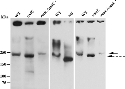
FIG. 4.
Characterization of EmaA in the O-PS mutants. Equivalent amounts of membrane protein from each strain was prepared and separated by electrophoresis using 4 to 15% gradient polyacrylamide Tris-HCl gels. The proteins were transferred to nitrocellulose and probed with a monoclonal antibody specific for EmaA. The solid arrow indicates the electrophoretic mobility of the EmaA monomers associated the wild type and complemented strains in the separating gel. The dashed arrow corresponds to the mobility of the EmaA monomers associated with the rmlC mutant strain in the separating gel. The immunoreactive material at the top of the immunoblot corresponds to EmaA aggregates associated with the stacking gel. WT, wild type (VT1169); rmlC, rhamnose epimerase mutant; rmlC/rlmC+, rhamnose epimerase complemented; wzt, ABC sugar transport mutant; waaL, O-antigen ligase mutant; waaL/waaL+, O-antigen ligase complemented.

