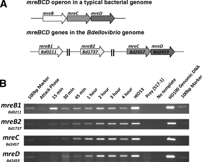
FIG. 2.
mreBCD gene organization and expression in B. bacteriovorus. (A) Schematic drawing of the mreBCD locus in a typical bacterial genome versus that of the B. bacteriovorus genome strain HD100. (B) Semiquantitative RT-PCR study of B. bacteriovorus mreBCD expression across a predatory infection cycle. RT-PCR reactions were carried out on RNA extracted at the time points across a synchronous lysate of B. bacteriovorus on the E. coli strain S17-1. Time points at which the RNA sample was taken are labeled on the figure. The HID13 strain used to assay HI gene expression. E. coli S17-1 RNA and no-template reactions provide negative controls, and HD100 genomic DNA was used as a template for a positive control. Primers for gene detection were designed to amplify an internal region of each mreBCD gene. The 100-bp marker from the NEB 100-bp ladder is visible in each marker lane.

