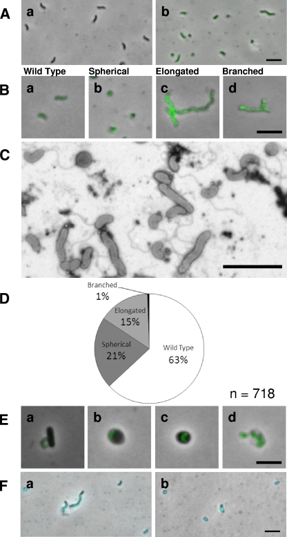
FIG. 5.
Analysis of the attack-phase morphologies of HD100 MreB2-mTFP strain. (A) Fluorescence images of the H100ABC, nonfluorescent “wild-type” control strain (a) and HD100 MreB2-mTFP attack-phase cells (b). (B) Attack-phase morphologies of MreB2-mTFP strain wild-type (a), spherical (b), elongated (c), and branched (d) cells. (C) Representative electron micrograph showing MreB2-mTFP attack-phase cells. The cells were stained with 0.5% uranyl acetate. (D) Pie chart representing a survey of 718 HD100 MreB2-mTFP attack-phase cell morphologies. (E) Fluorescence images of MreB2-mTFP strain at the attachment (a), early (b), and late (c) bdelloplast and lysis (d) stages of the HD predatory life cycle. (F) DAPI-stained fluorescence images of MreB2-mTFP attack-phase cells. Scale bar, 3 μm.

