
Figure 3.
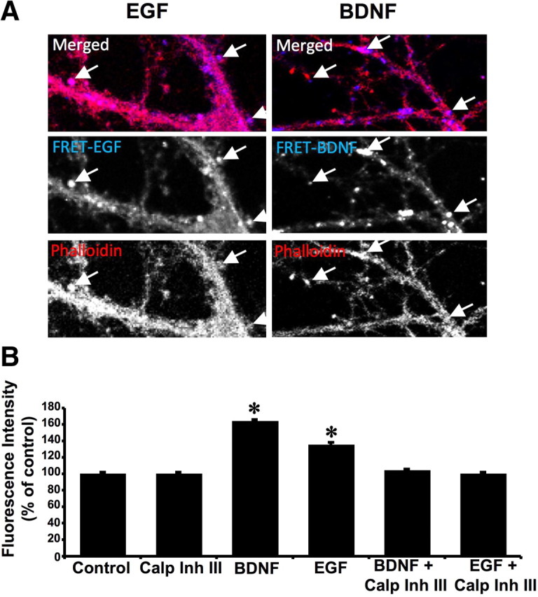
Calpain activation is localized in dendritic spine-like structures and stimulates actin polymerization. A, Hippocampal neurons were preloaded with the FRET substrate and treated with BDNF (50 ng/ml) for 10 min or EGF (20 ng/ml) for 2 min. They were then fixed, stained with phalloidin–Alexa Fluor594, and processed for confocal microscopy. Decrease in FRET signal (increased fluorescence) was observed in dendrites as well as in dendritic spine-like structures labeled with phalloidin–Alexa Fluor594 (arrows). B, Hippocampal neuronal cultures were pretreated with calpain inhibitor III (10 μm) for 20 min before adding vehicle or BDNF (50 ng/ml) for 20 min. At the end of incubation, actin polymerization was analyzed as described under Materials and Methods with the rhodamine–phalloidin fluorescence enhancement assay. Results of the rhodamine– phalloidin fluorescence were normalized (by subtracting control fluorescence signals) and represent means ± SEM of five experiments. *p < 0.001 (ANOVA, followed by Bonferroni's test).