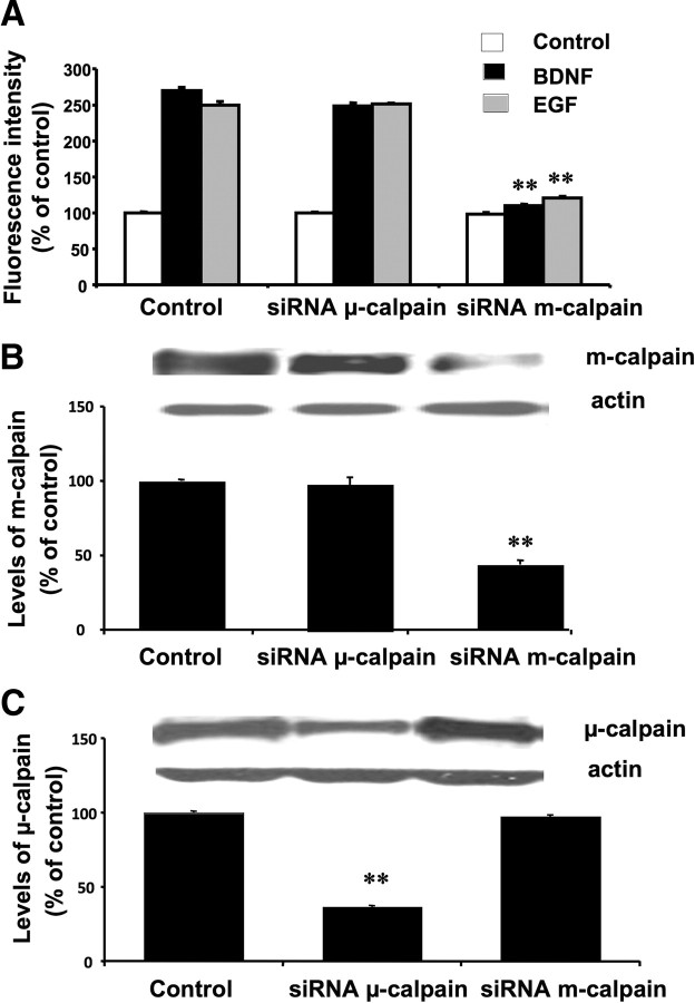
Figure 6.
Effect of siRNA against μ- or m-calpain on calpain activity in cultured cortical neurons. Cultured cortical neurons (DIV14) were transfected with 40 μm siRNA diluted in Neurobasal media and 40 μl of HiPerfect, as described in Materials and Methods. After 5 d, cells were preincubated with the FRET reagent for 3 h. They were then treated with vehicle (control), EGF (20 μg/ml), or BDNF (50 μg/ml) for 30 min. Cells were lysed, and fluorescence in the lysates was determined (A). Aliquots of cell lysates were also processed for Western blots with m-calpain (B) or μ-calpain (C) antibodies (actin antibodies were used as loading control). Fluorescence intensity results are expressed as percentage of values found in control-treated nontransfected neurons and are means ± SEM of three experiments. **p < 0.01 compared with the respective control values. Levels of m- and μ-calpain were normalized and expressed as percentage of values found in control-treated nontransfected neurons and are means ± SEM of nine experiments. **p < 0.01 compared with control values.

