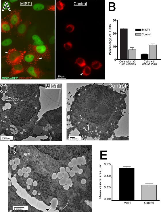
FIG. 6.
HGC-MIST1 cells form large exocrine granules upon PGC-RFP transfection. (A) Fluorescence microscopy image of HGC-27 cells stably expressing MIST1-eGFP (green nuclear staining) showing large PGC vesicles (arrowheads) that do not form in control cells (stably expressing eGFP alone) upon transfection with PGC-RFP. (B) The fraction of cells with multiple (≥3) large (≥1-μm) vesicles and diffuse, bright vesicles is quantified across multiple experiments (note that the remaining cells showed above-background RFP but were too dim to categorize). (C) TEM image of ZCs from HGC-MIST1 and control cells. Note the reduced size of secretory vesicles in the (control) HGC-27 cells. SV, secretory vesicle. (D) TEM image showing exocytosis of vesicle contents (arrowhead) in an HGC-MIST1 cell. (E) Vesicle sizes were quantified from TEM of multiple HGC-MIST1 and control cells.

