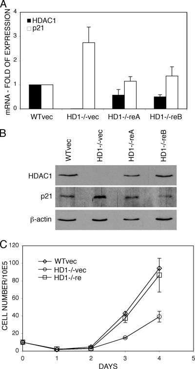
FIG. 1.
Reintroduction of HDAC1 into HDAC1−/− ES cells restores proliferation and low p21 expression. (A) Analysis of HDAC1 and p21 mRNA expression in ES cells. RNA from vector transfected HDAC1+/+ ES cells (WTvec) and HDAC1−/− ES cells (HD1−/−vec) and two different HDAC1 reintroduced ES cell lines (HD1−/−reA and HD1−/−reB) was analyzed by real-time PCR. Expression of HDAC1 and p21 is shown relative to β-actin. The relative expression in vector-transfected HDAC1+/+ cells was arbitrarily set to 1. The data are shown as mean values of three independent experiments with standard deviation (SD). (B) Immunoblot analysis of HDAC1 and p21 protein expression. Whole-cell extracts were prepared from vector-transfected HDAC1+/+ and HDAC1−/− ES cells and HDAC1 reintroduced ES cell lines (HD1−/−reA and HD1−/−reB) and analyzed for the presence of HDAC1 and p21. β-Actin was used as loading control. (C) Growth curves of vector-transfected HDAC1+/+ and HDAC1−/− ES cells and HDAC1−/− ES cells transfected with pMSCV-HDAC1 (HD1−/−re). For each cell line, 105 cells were seeded in triplicates, and aliquots were counted daily for 4 days and presented as mean values ± the SD.

