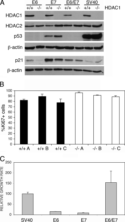
FIG. 4.
Transfection with viral oncogenes can rescue the proliferation/immortalization phenotype of primary HDAC1−/− MEFs. (A) Immunoblot analysis of HDAC1, HDAC2, p53, and p21 protein expression. Whole-cell extracts were prepared from HDAC1+/+ and HDAC1−/− MEF single clones obtained after transfection with HPV16 E6, HPV16 E7, HPV16 E6/E7, and SV40 LT. β-actin was used as loading control. (B) Proliferation of transformed HDAC+/+ and HDAC1−/− fibroblasts. SV40 LT-transformed HDAC+/+ and HDAC1−/− fibroblast lines were analyzed by indirect immunofluorescence microscopy for both HDAC1 and Ki67 expression. The fraction of Ki67-positive cells is depicted as the mean value ± the SD of triplicates for three independent cell lines of each genotype. (C) Relative proliferation rates of E6, E7, E6/E7, and SV40 LT-transfected MEFs. A total of 105 cells of at least three different single clones for each transfection were seeded and counted after time period of 3 days. Proliferation rates were determined and are presented as cell numbers per day relative to the value of SV40 LT cell lines in percentages as mean values ± the SD.

