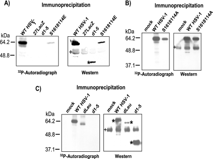
FIG. 4.
In vivo phosphorylation of wild-type HSV-1 KOS and phosphorylation site mutants. (A) HeLa cells were infected with KOS, 27-LacZ, S16,18,114E, and d1-5 at an MOI of 5. Cells were labeled with [32P]orthophosphate added 45 min after infection, and cells were harvested at 8 h after infection. Whole-cell lysates were immunoprecipitated with anti-ICP27 antibodies. Samples were fractionated by SDS-PAGE and transferred to nitrocellulose. The autoradiograph of the blot is shown in the left panel. After the decay of the radiolabel, the same blot was probed with antibody to ICP27 and is shown in the right panel. The asterisk marks the position of d1-5 protein. The bands denoted by the plus sign represent heavy-chain IgG from the immunoprecipitation. (B) HeLa cells were mock infected or were infected with KOS or S16,18,114A at an MOI of 5. In vivo labeling and Western blot analysis was performed as described for panel A. (C) HeLa cells were mock infected or were infected with HSV-1 KOS, dLeu, or d1-5 at an MOI of 5. Labeling and Western blot analysis were performed as described for panel A. The asterisks mark the position of wild-type and mutant ICP27. The bands denoted by the plus sign represent heavy-chain IgG from the immunoprecipitation.

