FIG. 7.
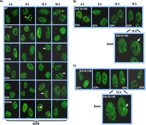
ICP4-containing replication compartment formation is reduced in ICP27 phosphorylation site mutant infections. (A) HeLa cells were infected with wild-type KOS, 27-LacZ, S16A, S18A, S114A, and S16,18A at an MOI of 5. Cells were fixed and stained with anti-ICP4 antibody at 4, 8, 12, and 16 h after infection. Yellow arrows point to ICP4-containing globular transcription/replication compartments in wild-type KOS-infected cells at 8 and 16 h after infection. White arrows point to ICP4 ring-like structures seen in the phosphorylation mutant infections. (B) HeLa cells were infected as described for panel A with S16,18,114A, and cells were fixed and stained at the times indicated in the figure. The image at 16 h after infection has been zoomed to view the ICP4 ring-like structures denoted by the white arrow. (C) Cells were infected with S16,18,114E as described for panel A. The image at 12 h has been zoomed to better demonstrate the ICP4 ring-like structures. The white arrow points to a ring-like structure.

