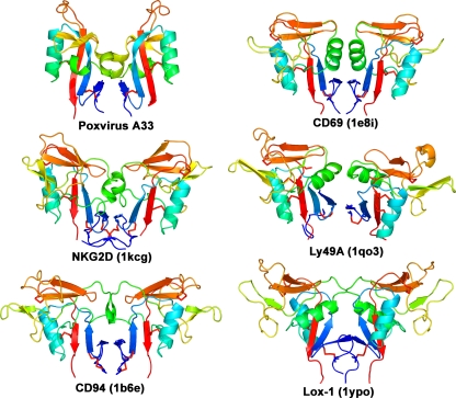
FIG. 3.
The A33 homodimer of CTLDs is structurally similar to CTLD dimers found in only a few other proteins. A ribbon depiction of the A33 dimer (Poxvirus A33) is shown at the upper left. Other known homodimers of CTLDs are from the immune system and are colored similarly for comparison: CD69 (PDB code 1e8i [46]), NKG2D (1kcg [59]), Ly49A (1qo3 [71]), CD94 (1b6e [5]), and Lox-1 (1ypo [56]). The KLRG1 dimer (3ff8 [45]) is also similar (not shown). The molecules are rainbow colored in a gradient from the N terminus (dark blue) to the C terminus (red). Disulfide bonds are shown in red. All proteins in this figure are type II transmembrane proteins.

