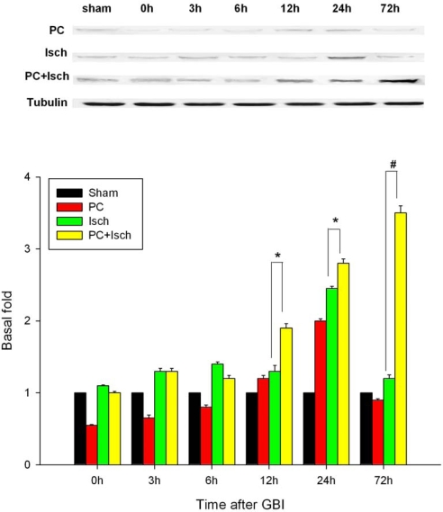
Figure 4.
Western blotting analysis of ASIC 2a activations and expressions in hippocampal CA1 regions.
Animals were subjected to preconditioning (PC), global ischemia (Isch), or preconditioning followed by ischemia (PC + Isch), by the four-vessel occlusion paradigm (PC, 3 min, 48 h before Isch, 15 min), followed by reperfusion. Rats were decapitated at 0 h, 3 h, 6 h, 12 h, 24 h or 72 h after reperfusion. Extracts from the hippocampi of the rats and sham controls were subjected to Western blotting with anti-ASIC 2a protein. Data are mean ± S.D. (n = 5). * p < 0.05, # p < 0.01 by nonparametric ANOVA followed by Dunn’s analysis comparing with ischemic rats.

