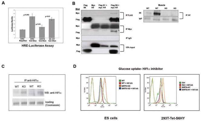
Figure 4. SIRT6 is a co-repressor of Hif1α.

(A) A luciferase reporter gene under the regulation of 3 tandem copies of Hypoxia-Responsive Elements (HRE) was co-transfected with empty vector (CMV), SIRT6 (S6) or SIRT6-HY (catalytic dead) plasmids into 293T cells, and subjected to low-glucose (5mM) conditions for 24 hr. Extracts were analyzed for Luciferase activity. (B) Left panel: A Flag control, a SIRT6-Flag or a SIRT1-Flag proteins were either expressed alone or co-expressed with Hif1α-Myc in 293T cells, and following immunoprecipitation (IP) with either a Flag, a Myc, or an IgG antibody, extracts were analyzed by Western blot, and probed with the indicated antibodies. Right Panel: lysates were prepared from SIRT6 WT and KO muscle, and following IP with anti-Hif1α antibody, extracts were analyzed by western blot probed with anti-SIRT6 antibody. The IgG band is shown as loading control. (C) Lysates were prepared from SIRT6 WT or KO ES cells, followed by IP and western blot with a Hif1α antibody. (D) ES cells (left panel) or 293T cells stably expressing a tetracycline inducible SIRT6 dominant negative allele (S6HY)(right panel) were treated with or without the Hif1α inhibitor #77 (Zimmer et al., 2008) and glucose uptake was measured by FACS, following 1 hr. exposure to NBDG. See also Figure S5.
