
Figure 7. Hif1α-dependent increased glycolysis and lactate production in SIRT6 deficient mice.
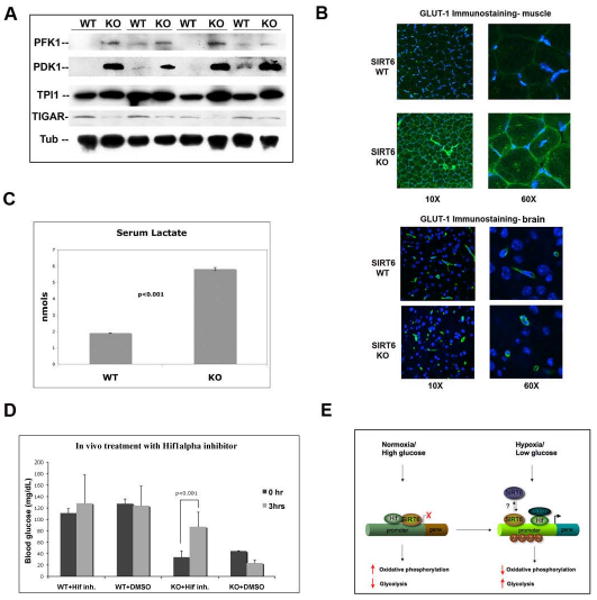
(A) Lysates were prepared from muscles of 4 littermate-pairs of SIRT6 WT and KO mice. Western analysis was carried out with antibodies against the indicated proteins. Tubulin was used as a loading control. (B) Immunostaining with a GLUT1 antibody (green) was carried out on muscles and brain from SIRT6 WT and KO mice. Nuclei were stained with DAPI (blue). Images were taken using a confocal microscope with constant laser beam for all images (KR: 39.8; IRIS: 2.0). (C) Serum was purified from SIRT6 WT and KO mice, and lactate was measured using the Lactate Assay Kit (BioVision). Error bars indicate the standard error of the mean. n=4 for each genotype. (D) A Hif1α small molecule inhibitor rescues the glucose phenotype in SIRT6 deficient mice. Hif1α inhibitor #77 (20μg/g weight) was injected i.p. in 19 days-old wild type and SIRT6 KO mice, and 30 min later blood was withdrawn for glucose measurement. 5% DMSO (dilution solution) was injected as control. (E) Model. Under normal nutrient conditions, SIRT6 inhibits expression of glycolytic genes acting as an histone deacetylase to co-repress Hif1α. This maintains proper flux of glucose to the TCA cycle. Under conditions of nutrient stress, SIRT6 is inactivated, allowing activation of Hif1α, recruitment of p300, acetylation of H3K9 at the promoters and increased expression of multiple metabolic genes, causing increased glycolysis and decreased mitochondrial respiration. See also Figure S7.