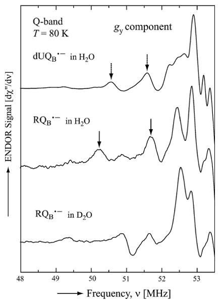
Fig. 4.

ENDOR powder spectra of and in quintuple mutant RCs. Arrows indicate the couplings between exchangeable protein protons that form H-bonds with (upper trace) and proposed to form H-bonds with (middle trace). Assignment of these latter peaks to H-bonds is supported by their absence upon exchange into D2O (lower trace). Spectra recorded at the magnetic field position corresponding to gy (see Fig. 3). Experimental conditions: T = 80 K, MW frequency = 35.03 GHz, MW power = 3 × 10⁻⁶ W, frequency modulation (FM) = ±140 kHz at a rate of 947 Hz. Number of scans: 18,000 (top), 500 (middle) and 2,600 (bottom). Scan time: 4 s
