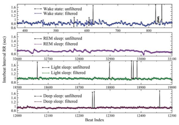
Fig. 1.
Representative recordings of segments of 500 RR intervals between consecutive normal heartbeats for a healthy young subject during deep sleep, light sleep, rapid eye movement (REM) sleep, and an intermediate wake state (from bottom to top), part of an 8 h polysomnographic recording. A small percentage of artefacts (e.g., spikes marked with ×) have been filtered before the actual analysis (see Section II). Data show ×(a) gradual decrease of the standard deviation, σRR (also denoted as SDNN) from wake to REM, to light, to deep sleep (see Fig. 2), and (b) that the heartbeat signal is more homogeneous in deep sleep compared to light sleep, REM sleep, and wake where it exhibits more irregular fluctuations (see Fig. 3).

