
Figure 6.
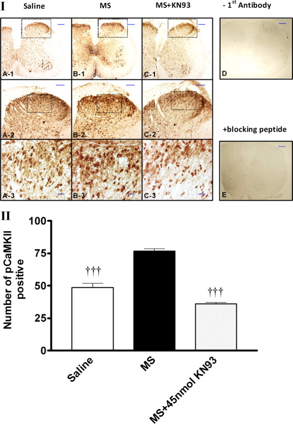
Immunohistochemical staining of pCaMKIIα expression after the treatment with KN93. I, Morphine- or saline-treated mice were administered (intrathecally) with KN93 (15–45 nmol), KN92 (45 nmol), or saline on day 5. Mice were killed 1 h after the treatment with saline or KN93 (45 nmol, i.t.). The lumbar spinal section was dissected out and immunostained with pCaMKIIα antibody. II, Quantitative analysis of pCaMKIIα immunoreactivity was performed by counting the number of positively stained cells using the MetaMorph Imaging Software. No pCaMKIIα immunoreactivity was detected if the first antibody was omitted (D) or if the first antibody was incubated in the presence of pCaMKIIαT286 blocking peptide (Santa Cruz Biotechnology). Data are expressed as mean ± SEM, †††p < 0.001, compared with the morphine-treated group; n = 3 for each group. Scale bars are 200 μm (A-1, B-1, C-1, D, E), 100 μm (A-2, B-2, C-2), or 20 μm (A-3, B-3, C-3).