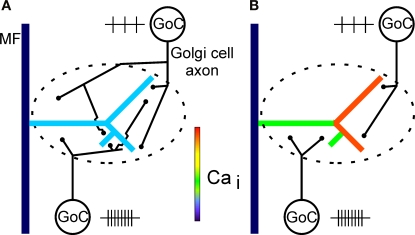
Figure 8.
Diagram of hypothetical Golgi cell innervation patterns in a glomerulus with a complex mossy fibre rosette. (A) In one possible scenario, Golgi cells (GoC) innervating the same glomerulus overlap in their axonal innervation territory, making a large glomerulus a functional entity. The global presynaptic inhibition of the rosette is due to summation of activity of all Golgi cells innervating the glomerulus. Control situation for (A) and (B) with no Golgi cell inhibition is indicated in Figure 7G. (B) In another scenario, no or marginal overlap of Golgi cell axons occur within a large glomerulus. Different compartments of a large complex rosette could in this way be subject to local presynaptic inhibition by different Golgi cell populations. This indicates that control of input to the cerebellar cortex could be more fine-grained than suggested by the scenario in (A), where an entire glomerulus functions as an input unit. Future anatomical analysis may allow to differentiate between these alternatives. In (B), the relative activity levels (indicated by the number and spacing of small vertical lines close to each cell) of Golgi cells may dynamically control presynaptic calcium influx in different branches of a rosette. Feedback as well as lateral-inhibition by Golgi cells could functionally compartmentalize the glomerulus. Note that the Golgi cells in the diagrams (A) and (B) represent two different cell populations and are not an indication of the actual number of Golgi cells innervating a glomerulus. Note also that the innervation scenarios depicted in (A) and (B) could be considered to illustrate opposite ends of a spectrum.

