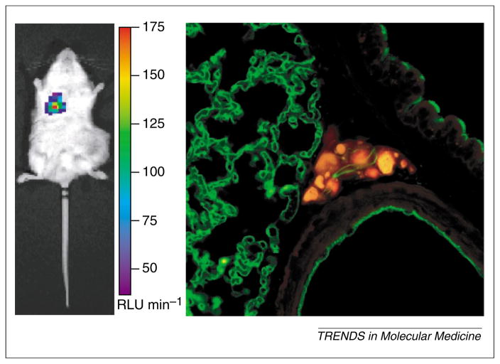
Figure 4.
Verification of positive optical signals. Animals bearing prostate tumor received 3.6 or 7.2 × 10⁷ infectious units of prostate-targeted adenovirus, AdPSE-BC-FL, administered via the tail vein. (a) Histological analysis to assess positive signal. Twelve days after administration, an optical signal was observed in the lung (left panel) of an animal that received 3.6 × 10⁷ infectious units of virus. The positive optical signal in this animal was correlated with the presence of metastatic human cancer cells in the lung (right panel). Cancer cells in the lung sections were visualized by confocal microscopy using CY-3 conjugated (red) human-specific pan-cytokeratin antibody (BioGenex Laboratories). Lung blood vessels were visualized by FITC-lectin (green).

