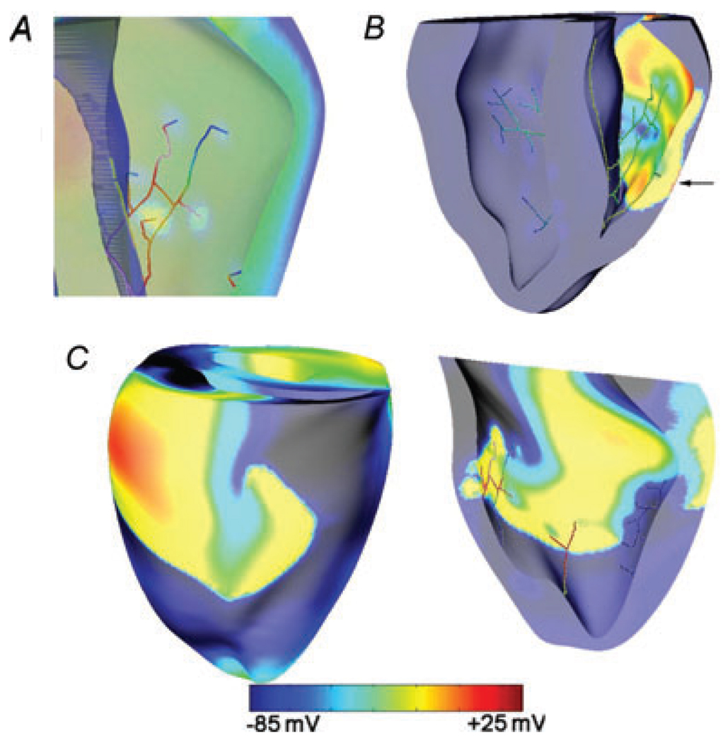
Figure 7. Post-shock Purkinje system activation determines isoelectric window.
A, the earliest endocardial activations following the 3.6 V cm−1 shock during the burst pacing protocol (BCL 130 ms) are induced by Purkinje strands. B, these activations propagate transmurally to appear as the first propagated postshock breakthrough on the epicardium that marked the end of IW. Black arrow shows the breakthrough location. C, postshock activations degrade into re-entry; rotors are seen on RV epicardium (left) and endocardium (right).

