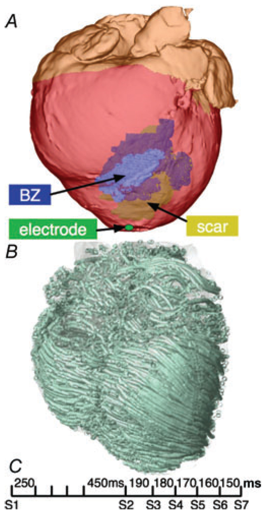
Figure 8. MRI-derived infarcted canine heart model.
A, anterior view of the infarcted canine heart with inexcitable scar (yellow) and partly viable border zone (BZ; blue). B, the corresponding fibre orientation calculated from the DTMR, visualized as streamlines. C, aggressive pacing protocol delivered at the LV apex to induce VT.

