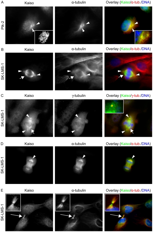
Figure 3. Kaiso localizes at centrosomes and spindle microtubules during mitosis.
(A) Staining for Kaiso and α-tubulin in Ptk-2 cells during prophase. Kaiso accumulates at the centrosomal region (arrowheads). The left inset shows chromosomal condensation, while the right inset shows in more detail the colocalization of Kaiso and α-tubulin. During metaphase (B, C) Kaiso is also present on the entire spindle (short arrow). (B) Staining for Kaiso and α-tubulin in SK-LMS-1 cells; photographs were taken while focusing on the spindle microtubules. (C) Localization pattern of Kaiso in SK-LMS-1 cells compared to that of γ-tubulin at the centrosomes; photographs were taken by focusing on centrosomes. The inset magnifies the upper centrosomal region. (D) During anaphase of SK-LMS-1 cells Kaiso localizes at the microtubules and centrosomes, and during cytokinesis (E) it localizes at the midbody (long arrow); insets show magnifications of the midbody region. Monoclonal antibodies were used to detect Kaiso (mouse mAb 6F) or α-tubulin (rat mAb YOL1/34). Secondary antibodies were an Alexa 488-conjugated anti-mouse antibody for detection of Kaiso, and an Alexa 594-conjugated anti-rat antibody for α-tubulin. For γ-tubulin detection, a rabbit polyclonal antibody was used, followed by an Alexa 594-conjugated anti-rabbit secondary antibody. DNA was stained with DAPI. Cells were imaged (100× objective lens) with a Zeiss Axiophot microscope.

