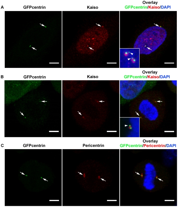
Figure 4. Kaiso localizes in the PCM.
Fluorescence microscopy of methanol-fixed HeLa-GFP-centrin cells in either G2/M phase (A) or metaphase (B, C). In (A) and (B) Kaiso was immunolabeled with pAb S1337; in (C) pericentrin was detected by pAb ab4448. Alexa 594-conjugated anti-rabbit secondary antibody was used in all cases and DNA was stained with DAPI. Cells were imaged with a Leica SP5 confocal microscope. Each figure is a projection of a confocal image stack. Colocalization of centrin with Kaiso and pericentrin was confirmed in layers of 0.77 mm thickness. Scale bar, 5 µm. Arrows point at centrosomes. Insets in (A) and (B) show magnified overlay pictures of centriolar regions, each time in a second cell of the same slides. The region of Kaiso-positive staining is broader than the centrin-positive centrioles and overlaps with the mother centriole rather than the daughter centriole (arrowheads). Spindle-associated Kaiso was not obvious in the present experiments, as other fixation and staining conditions were used.

