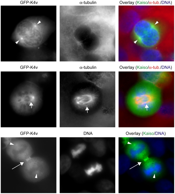
Figure 7. The GFP-tagged Kaiso fragment K4v, comprising SA1, localizes at the spindle material during mitosis.
HEK293 cells were transfected with construct GFP-K4v (see also Fig. 1). During the G2/M phase (upper row), GFP-K4v concentrates at the centrosomes (arrowheads). In metaphase (middle row), it is visible along the entire spindle (short arrow). During cytokinesis (lower row), it is present in the midbody (long arrow) and the centrosomes (arrowheads). Rat monoclonal antibody YOL1/34 was used to detect α-tubulin. Secondary antibody was an Alexa 594-conjugated anti-rat antibody. DNA was stained with DAPI. Cells were imaged (100× objective lens) with a Zeiss Axiophot microscope.

