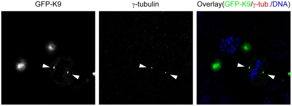
Figure 8. The GFP-tagged Kaiso fragment K9, comprising SA2, localizes at the centrosomes during mitosis.
HEK293 cells were transfected with construct GFP-K9 (see also Fig. 1). This fragment localizes only at the centrosomes (arrowheads), which were visualized with a polyclonal anti-γ-tubulin antibody followed by an Alexa 594-conjugated anti-rabbit secondary antibody. DNA was stained with DAPI and cells were imaged (63× objective lens) with a Leica DM IRE2 microscope.

